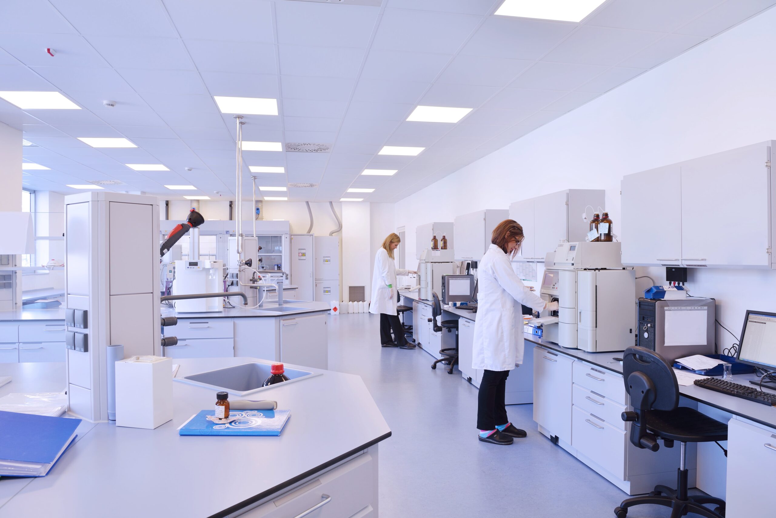
In the ever-evolving landscape of pathology, automation has emerged as a transformative force, revolutionizing traditional laboratory practices.
One of the most significant advancements in this domain is the automated immunohistochemistry (IHC), a technology that has streamlined high-throughput analysis in pathology, leading to quicker and more accurate diagnoses.
Central to this innovation is the integration of cutting-edge tools like the Process Record Slide (PRS) tool, which stands as a beacon of quality assurance, enhancing the reliability and validity of immunohistochemistry staining assays.
The Rise of Automated Immunohistochemistry:
Traditional IHC staining methods often encounter challenges in terms of consistency, especially when dealing with a large volume of samples, where the pathologist will need to prepare multiple stains on the tissue for investigation.
Automation has tackled these issues head-on by introducing precision and efficiency previously unseen in pathology laboratories. Automated immunohistochemistry systems now allow for the simultaneous processing of multiple samples, drastically reducing turnaround times and improving diagnostic accuracy.

PRS Landing Med Automated Slide Staining Machine
Steps to Operate an Automated Immunohistochemistry Equipment
There are about ten steps that are involved when operating an automated immunohistochemistry process.
- Sample Preparation:
Prepare tissue sections on glass slides following standard histology procedures. Ensure that the tissue sections are properly fixed and processed before staining.
- Loading Samples
Place the prepared slides onto the slide rack or carousel of the Landing Med Automated Slide Staining Machine, automated IHC instrument by Process Record Slide Limited. Ensure proper labeling and organization to avoid confusion during the staining process.
- Reagent Preparation
Following the manufacturer’s instructions, prepare the necessary reagents, including antibodies, detection systems, and buffers. Ensure that all reagents are at the appropriate temperature and are properly mixed.
- Loading Reagents
Load the required reagents into their respective compartments or containers within the automated staining instrument. Some systems have specific slots for primary antibodies, secondary antibodies, and other solutions.
- Program Selection:
Choose the appropriate staining protocol or program from the system’s menu. Automated IHC instruments like the Landing Med Automated Slide Staining Machine have pre-programmed protocols for various antibodies and staining procedures. Select the one that corresponds to your specific staining requirements.
- Start the Automated Staining Process:
Initiate the staining process by starting the automated instrument. The system will proceed with the staining steps, including deparaffinization, antigen retrieval, blocking, incubation with primary and secondary antibodies, chromogen reaction, and counterstaining.
- Monitoring and Troubleshooting:
Monitor the staining process to ensure that it proceeds smoothly. Automated systems like Landing Med Automated Slide Staining Machine have real-time monitoring features. In case of any issues or errors, the system may provide alerts. Troubleshoot problems promptly, following the instrument’s user manual or contacting technical support if necessary.
- Post-Staining Steps:
Once the staining process is complete, remove the slides from the instrument. Perform any necessary post-staining steps, such as washing and mounting coverslips. Allow the slides to dry properly before examination under a microscope.
- Quality Control and Validation:
Conduct quality control checks to ensure the accuracy and reliability of the staining. Use appropriate positive and negative controls, including non-tissue controls like the Process Record Slide (PRS) tool, to validate the staining results.
- Result Interpretation:
Examine the stained slides under a microscope. Evaluate the staining patterns and intensity, comparing them with established criteria for the specific antibody or biomarker. Document and interpret the results accurately for further analysis and reporting.
The Role of Process Record Slide (PRS) Tool in Automated Immunohistochemistry
The Process Record Slide (PRS) tool serves as a pivotal quality indicator, enhancing the reliability and accuracy of the immunohistochemistry (IHC) staining process. Every laboratory assay necessitates the use of controls, acting as benchmarks to ensure the delivery of accurate results.
The PRS tool stands out as an evidence-based quality control instrument, capable of identifying errors during staining, particularly in antigen retrieval or when using expired or substandard staining materials. These controls are indispensable for instilling confidence in doctors regarding the results and preventing the occurrence of false positives or negatives.
Unlike tissue-based controls, non-tissue controls, such as the PRS tool, are not limited by ethical considerations. This distinction allows for their broader application.
Moreover, they overcome the challenges related to the reliability and reproducibility associated with tissue-based controls. Non-tissue controls significantly contribute to enhancing the efficiency of automated immunohistochemistry, making them a valuable asset in the realm of laboratory diagnostics.
Increasing Quality, Reliability, and Validity:
The integration of the PRS tool in automated immunohistochemistry brings a multitude of benefits.
Firstly, it dramatically increases the overall quality of staining procedures. By providing a standardized protocol. It minimizes human errors and ensures consistency across various samples, even in high-throughput scenarios. This standardization significantly reduces the variability that might arise due to different technicians or laboratories, thus enhancing the reliability of results.
Secondly, the PRS tool amplifies the reliability of immunohistochemistry assays. With its ability to maintain a consistent staining protocol, it minimizes the chances of false positives or false negatives. This reliability is paramount, especially in cancer diagnosis, where accurate identification of specific biomarkers can dictate treatment decisions and prognoses.
Moreover, the PRS tool elevates the validity of immunohistochemistry assays. By offering a detailed record of the staining process, it ensures that the results are not only accurate but also credible. Pathologists and researchers can have confidence in the outcomes, knowing that a standardized and documented procedure backs them.
Conclusion:
Automated immunohistochemistry, coupled with the implementation of tools like the Process Record Slide (PRS) tool, has ushered in a new era of efficiency, accuracy, and reliability in pathology laboratories.
The ability to conduct high-throughput analysis while maintaining the highest standards of quality transforms how diseases are diagnosed and understood.
As technology advances, integrating automated systems and quality indicator tools like PRS will undoubtedly play a pivotal role in shaping the future of pathology, ensuring that patients receive the most accurate and timely diagnoses possible.


